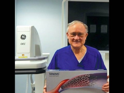
By Ronald Bush, MD, FACS
In 1992, I left a busy practice of thoracic and vascular surgery and began the in-office treatment of varicose veins. At that time, high ligation and stripping using tumescent with ultra sound guided assistance was our treatment of choice for saphenous insufficiency.
Since that time, many changes have occurred in the treatment of venous disease including, but not limited to, lasers, radio frequency, foam sclerotherapy, glue and numerous other devices for the treatment of deep venous pathology.
I have been associated with many of the vein conferences over the years. However, most of these conferences lack specific information on how to deal with the many aspects of venous disease. About 80 percent of all patients can be classified as cosmetic even if they have saphenous insufficiency. Many times, the insufficiency is secondary to the cosmetic issue.
Most patients present with either spider veins or enlarged veins in other areas of the body such as the hand and face. Very little is written about the pathophysiology of venous disease manifested at the skin level.
All stages of venous disease can be evident from just examining the skin. The skin, being the largest organ of the body, contains clues to abnormalities located in the systemic and venous system.
Think of this concept as follows:
- Class I – Spider veins are the result of transmitted pressure (Cutaneous Venous Hypertension from a deeper source). Based on our extensive histological research, all telangiectasia occurs in the reticular dermis. The average depth is 700 microns.
- Class II – Class ll disease is actually a manifestation of varicosities, which are subdermal.
- Class III – Class lll disease is manifested as edema or swelling of the dermal and subdermal tissue.
- Class IV-V – Class IV- V is manifested by changes in both the dermis and squamous layer.
So, in essence, all forms of venous disease are manifested by transmission of pressure to a small area that measures only 1.5mm in thickness.
Based on our extensive work in the dermatopathology lab at Water’s Edge Dermatology, we have been able to determine the effects of different modalities of treatment to various pathological venous conditions. We have also been able to incorporate micro-surgical techniques based on our extensive studies.
AESTHETIC VEIN CONFERENCE
Due to the lack of specific knowledge that most practitioners have in the treatment of numerous cosmetic issues of venous disease, we have developed the Aesthetic Vein Conference, an intensive course based on histology and anatomic principles that allow for safe and effective treatment for many vein related problems.
The next is Oct. 13. For additional information, call 407-900-8346 or see aestheticveintraining.com. Conference fee is $400 and is limited to the first 75 to register.
A few of the topics included in the conference include spider veins, varicose veins, hand veins, breast veins and, most importantly, veins of the face. Ultrasound, ohmic thermolysis, laser and sclerotherapy will be demonstrated in patients with complex problems.
In our clinic, we have treated more than 200 patients with dilated facial veins using foam sclerotherapy. Aesthetic Vein Conference will demonstrate new techniques as well as debunk many of the long-held tenants of the treatment of venous disease.
Our guidelines for treatment of facial veins are based on anatomic principles and foam containment. The greatest risk in using foam sclerotherapy on the face is not blindness, but superior ophthalmic vein or cavernous sinus thrombosis.
None of the established vein conferences discuss in detail the mechanism and possible complications of sclerotherapy for telangiectasia. Aesthetic Vein Conference provides details on cellular interaction in sclerotherapy as well techniques to avoid and treat complications.
Aesthetic Vein Conference goal is to provide a scientific basis for the treatment of venous disease in all locations and discuss techniques to provide rapid cosmetically superior results. The following images represent a challenge in treating spider veins. Spider vein aneurysm dilations is more prominent than you might think, and special techniques must be done to clear them up. VTN


